


Valgus foot deformation is most commonly innate. However, in some cases - with paralysis, traumatic lesions - can occur in the mature period of life. The main symptoms of pathology are pains in the field of foot and muscles of the lower leg, visible violation of the foot forms, as well as a change of walk. Diagnosis of the disease is carried out by clinical examination, X -Ray, electromyography, etc. Treatment includes conservative and surgical methods. However, proper efficiency is noticeable only in reconstructive operations.
What is this disease?
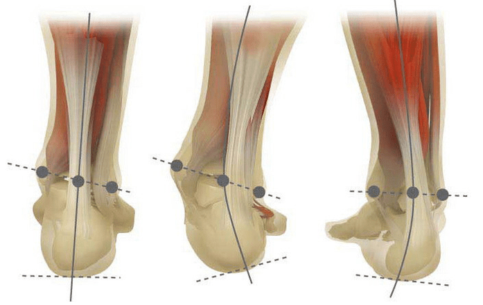
Valgus deformation is the curvature of the foot, characterized by the flattening of its longitudinal port. Usually the inner edge of the foot descends ("drops"), and the heel turned out.
The base of the person, thanks to his location, takes over the pressure of the whole mass of the human body. For this reason, it has a special anatomical structure, which enables depreciation, balancing and stabilization of the movement. However, an important component for the implementation of these tasks is an accurate stop.
Today, the most important problem is the traumatology and orthopedic valgus deformation of the foot. The meeting is estimated at 30-58%, where 2/3 cases are congregate disorders.
Pathology is largely socially significant, as it covers all age groups, and also helps in an insightful spinal column, early development of osteochondrosis and arthrosis of lower extremity.
When you take your legs (if you look at them from behind), at the level of deformation similar to the X-level: the ankles are in touch, while the heel is at a distance of 5-6 centimeters from each other.
Most often the pathology is innate into nature and is diagnosed with children in the hospital (or immediately after the start of walking). Similar condition adapts to 5 years, after which (in the absence of correct treatment) the child develops straight legs.
Why does it appear?
It is believed that the main reason for the effect of the foot deformation of the leg inadequate function of the rear teeper muscle or weakness of the ligamental apparatus.
Today, other factors are different in the development of pathology:
- Congenital abnormalities with the wrong location of bone legs or shortening the tendon (vertical ram bone, a short bunch of tendons);
- Holding disorders when the foot deformation is made up of the curvature of the spinal column;
- Traumatic lesions (Foot bone fractures, lower legs, hips or knees, ruptures of ligamental and tedative apparatus);
- Paralysis (immobilization) Due to damage to the nerve system for encephalitis, poliomyelitis, stroke, breach of cerebrospinal scrolls for hernias, etc. ;
- Spasm (constant contraction) of the lower leg muscle;
- Simultaneous diseases: Bone system with vitamin D (Rachit), diabetes Melitus, osteoporosis (bone density reduction), damaged thyroid glands and parathyroid glands, etc. ;
- Increased body weight, including a fast weight demand in menopausal or during pregnancy.

Development of pathology also facilitates irregularly selected shoes or excessive correction of childhood club.
Degree and stage of disease
The severity of pathology (power of manifestation) is divided with degrees:
- Light with the height of the vault of 1, 5-2 centimeters and the angle of the tilt of fifth to 15 degrees;
- Average, when the onion flattels in 1. Inches, and the angle decreases at 10 degrees;
- Heavy on the height of the vault to 0, 5 cm, and the angle of the heel slope is 5 degrees.
Depending on the inclusion of certain structures, the following phases of curvature are included:
- There are no bone deformations, pain is determined on the internal ankle surface (in the area of rear tibial muscle fastening);
- The curvature is light, the fifth is discarded a little;
- The foot was assigned, and the deformation is fixed (not correctly corrected);
- Wenting adheres not only in the foot, but also in the ankle joint.
Symptoms
In the first phase, patients have been disturbed periodic pain after a long walk or long vertical load (standing or sitting at the foot). As a rule, the pain syndrome intensifies when walking in irregularly selected shoes. The next phase of the disease is associated with the occurrence of foot curvature: patients in a standing position do not rely on the outer edge of the foot, but with an entire area. An easy change of Hot is noticed.

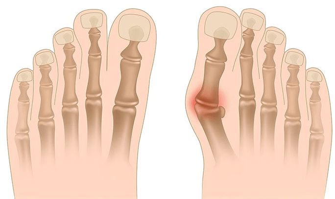
In the third phase, a protrusion of bone is determined (noticeably lower than the ankle on the internal ankle), as well as the power-out of the heel outside (the patient stands on the end of the heel edge). Advanced deformation of Valgus is characterized by pronounced curvature and the foot itself and the wrist of the ankle. Patients complain about heavy pain in lower leg muscles, as well as significant breach violations: knees are rubbed against each other, while the right and left foot is on some distance.
The severe curvature of the foot is often complicated by the deformation of the spinal column (of the pelvic shoulder and wing positions), osteochondrose (intervertised disk) (an intervertital disk of the creation) or arthrosis.
How to diagnose?
The diagnosis of the curvature of the foot consists of:
- Clinical inspection, during which the orthopediate detects the reduction of foot arches, the deviation of the bone of the heel and RAM, visible "disappearing" of the external and protrusion of internal ankles.
- X -RAY - Affordable and informative method you can specify a change in bone inclination and linear parameters. These indicators are required to execute the final diagnosis and clarification of deformation degrees.
- Method of registration of steps focused on determining the correct functional state of limb. The method consists in registration of the support time of individual feet parts when performing steps. During the study, the foot rolling phases are also studied, which reflects the balance of the lower limb muscles.
- Dynamic electromyography, which records the electrical activity of the studied muscles and its dependence on the steps of the steps.
- Photoplosmography with digital processing that allows you to get all standard indicators and determine the type / degree of curvature with great accuracy.
Additional neurologist counseling (with deformations due to cramps or paralysis), endocrinologists (in case of diabetes or thyroid / paratyroid gland) and gynecologists (when a threat is necessary). If the curvature of the foot appeared against the background of osteoporosis, densitometry is needed - a bone density study.
Treatment
Among the main methods of treatment of the curvature of valgus, conservative and operational and operational and operational. Do not destroy painful compounds in fat and injections!
Conservative approach
This type of assistance aims to solve the symptoms of the disease, but does not eliminate the root cause of pathology.
The technique includes:
- The use of orthopedic inspections for support and plus bones, onions, as well as the elimination of mid-mid-mid and the back of the foot;
- Taping - fastening of foot and ankle using special sticky ribbons with proper elasticity. The tape bar is used for about 3-5 days, after which they were replaced;
- sewing orthopedic shoes per individual standards;
- Use of orthosis and other feet and ankle fastening devices.
Conservative methods include physiotherapy procedures (ozokerites, paraffin applications, electrophoresis, magnetic effects), massage and complex of physiotherapy exercises, developed for a specific clinical case. Be careful! Today, most experts prefer surgical treatment methods, because the conservative therapy is inefficient (according to statistics, it is useless in 60% of cases).
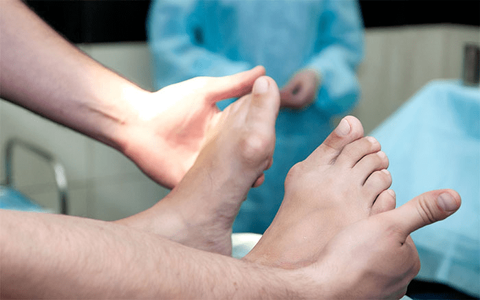
Surgical intervention
The scope of work and its type depends on the direct phase of the disease. Thus, the first degree of deformation valgus is treated with synonectomy (removal of a tetin shell for correction of general tension) or osteotomy (dissection) heel to return to an anatomical accurate position. In the second phase of the disease development, the transplant of the finger tendon is used. Such an intervention is usually done on the background of the heel or arthrodesis in the Ramn (surgical immobilization of the compound between RAM and scaphoid bones).
The curvature of the III of the degree requires arthrodesis of several foot joints at once: plus without cubic and desired shoulder. Such three-skonni immobilization is often complemented by the bonies of the heel bones. At the point of pathology, reconstructive operations are needed not only on foot, but also on ankle. In this case, the instability of the ligamental apparatus adapts to transplants (from one's own body or artificial material). The scope of operations on the foot is equal to the same as III degree of curvature.
Recovery period
Rehabilitation includes walking without support on the operated leg for 2 months. At the same time, the patient must wear removable gypsum long of 1, 5 to 3 months. Active operational leg movements are recommended to start after 1, 5 months after surgery. By 3. months, the complex of strengthening physical education is introduced. However, after that, patients are prohibited from lumpy activities of walking and active sports activities. It is worth noting that it is possible to judge the final result of surgery just six months later.
Preventive measures
The prevention of Valgus deformation includes the following measures:
- Early correction of innate anomalies with irregular layout of legs or shortening cereal tendons (vertical solvent bone, short toe);
- Correcting the disorders of holding (Scoliosis, etc. );
- Timely treatment of traumatic lesions (feet of feet, lower legs, thighs or wrist knees, rupture of the ligamental and tedious apparatus);
- Correct rehabilitation after paralysis (immobilization) for damage to the nerve system for encephalitis, poliomyelitis, stroke, breach of cerebrospinal roots for hernia, etc. ;
- Relief Grč (permanent reduction) muscle lower leg;
- Therapy for simultaneous diseases: bone systems with vitamin D (rikes), diabetes Melitus, osteoporosis (reduction of bone density), damaged thyroid function and parathyroid glands, etc. ;
- Reducing body weight to normal (especially with rapid gain in postmenface or due to pregnancy);
- Choice of orthopedic shoes or use of supineers;
- Moderate Club Correction without "hyper-correction" treatment leading to secondary secretion of the feet valgus.
Prevention of disease advancement is the use of conservative methods and early reconstructive operations. In this case, physical activity is limited to prevent the destruction and curvature of the ankle joints. Remember, timely treatment of the leg deformity does not only improve the quality of life of patients, but prevents the development of osteochondrose and arthrosis of knee or hook.